Figure 1.
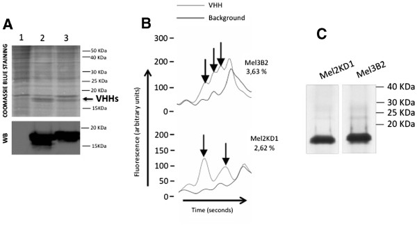
Expression of recombinant VHHs inT.nilarvae.A. SDS-PAGE analysis and Coomassie blue staining or Western blot (WB) of total soluble protein extracts of larvae infected with control baculovirus (lane 1), baculovirus expressing VHH Mel2KD1 (lane 2) or baculovirus expressing VHH Mel3B2 (lane 3). Western blot was carried out using an anti-VHH polyclonal antibody. The arrow indicates VHH bands. B. Analysis by capillary electrophoresis (Experion; Bio-Rad) of larvae extracts containing VHHs 3B2 and 2KD1. The arrows indicate the peaks used to determine the percentage of each recombinant VHH by subtracting the background proteins from the larvae extracts. C. SDS-PAGE analysis and Coomassie blue staining of recombinant VHHs purified by affinity chromatography.

