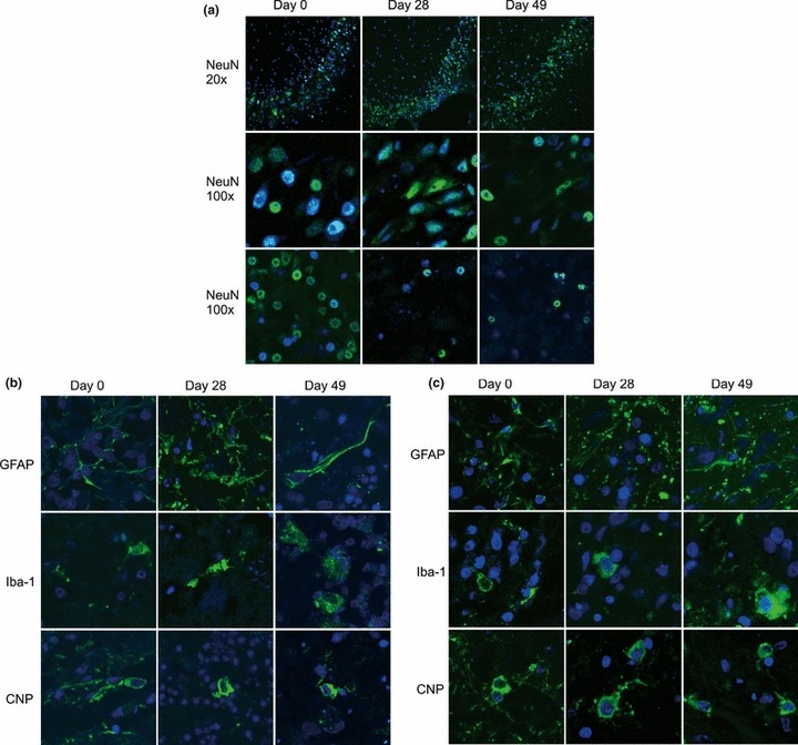
Figure 4.

(a) Double-immunofluorescence with antibodies specific for neurons (NeuN, green) on cryosections of hippocampal and cerebellar brain-slices of the same experiment as in Figure 3 at days 0, 28 and 49 in vitro. Nuclei were stained with TOTO-3 (blue). Top row: 20× magnification of the dentate gyrus of the hippocampus. Middle row: 100× magnification of the dentate gyrus of the hippocampus. Bottom row: 100× magnification of the granule cell layer in the cerebellar cortex. (b) Cryosections of hippocampal slices from the same experiment as in Figure 3 were stained (green) with antibodies specific for astrocytes (GFAP), microglia (Iba-1) or oligodendrocytes (CNP). Nuclei were stained with TOTO-3 (blue). Images are taken from the dentate gyrus. Magnification: 100×. (c) Cryosections of cerebellar brain-slice cultures from the same experiment as in Figure 3 were stained with the same antibodies as the hippocampal slices (days 0, 28 and 49 in vitro, 100× magnification). Images were digitally enhanced and gamma settings were adjusted using the FluoView software (Olympus FV10-ASW Version 01.07.01.00).
