
Figure 6.
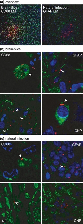
Comparison of infected brain-slices with natural infections. In all images, Listeria monocytogenes are stained in red (LM). Microglia (CD68), neuronal processes (NF), astrocytes (GFAP) and oligodendrocytes (CNP) are stained in green. Nuclei (TOTO-3) are blue. Images were digitally enhanced and gamma settings were adjusted using the FluoView software (Olympus FV10-ASW Version 01.07.01.00). (a) Overview: focal growth of L. monocytogenes (red) in brain-slices and natural cases of listeriosis. On the left microglia are stained in green (10× magnification). On the right astrocytes are stained in green (20× magnification). (b) Immunofluorescence on cryosections of infected brain-slices. CD68: L. monocytogenes are present in microglial cells (arrowhead, 300× magnification). Neurofilament (NF): L. monocytogenes are closely associated with axons (arrowheads, 200× magnification). GFAP: L. monocytogenes are closely associated with astrocytic processes (arrowhead, 200× magnification). CNP: in brain-slices, L. monocytogenes are only rarely found inside oligodendrocytes (arrowhead, 600× magnification). (c) Immunofluorescence on paraffin-sections of a natural case of listeric encephalitis. CD68: L. monocytogenes are present in microglial cells (arrowhead, 300× magnification). Neurofilament (NF): L. monocytogenes are closely associated with axons (arrowhead, 300× magnification). GFAP: L. monocytogenes are closely associated with astrocytic processes (arrowhead, 400× magnification). CNP: in natural infections no L. monocytogenes were found within oligodendrocytes (400× magnification).