
Figure 5.
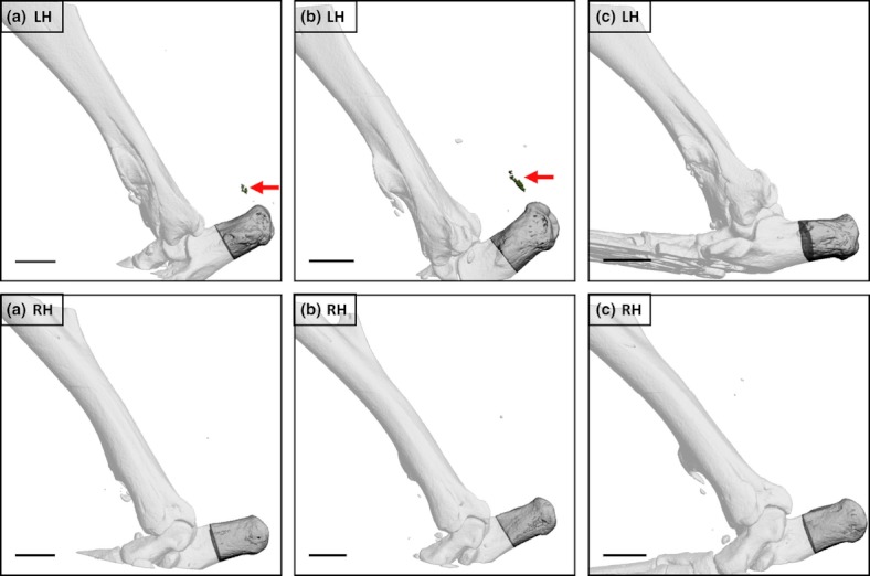
Left (LHs) and right (RHs) hindlimbs from three male C57Bl/6 mice 5 weeks following LH needle injury (performed when 10 weeks old). Arrows indicate mineralization close to the enthesis of the Achilles tendon within the LH in two of three individuals (a, b). A mineralized lesion was present in the same location in the third animal (c) but the density was too low for extraction using global thresholding protocols. No Achilles tendon-associated mineralization was detectable in the RH in any animal. Additional opacities are visible close to the Achilles tendon in some images but were positioned outside the skin or, in one case, had a far higher density than bone in that hindlimb and likely represented metallic debris deposited at the time of surgery. Bars = 1mm.