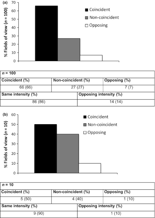
Figure 4.

EpCAM and MMP7 expression correlates in vivo in primary human samples. Serial cryosections of normal (n = 10) and head and neck carcinoma samples (n = 20) were stained with EpCAM- or MMP7-specific antibodies. Fields of view (FOV) of normal mucosa (one FOV/sample) and tumours (five FOV/sample) were assessed with respect to staining patterns and intensity of EpCAM and MMP7. Results are given as percentages of FOV with coincident, non-coincident or opposing patterns for tumours (a) and normal mucosas (b) Similarities in staining intensities are given in each table.
