Figure 6.

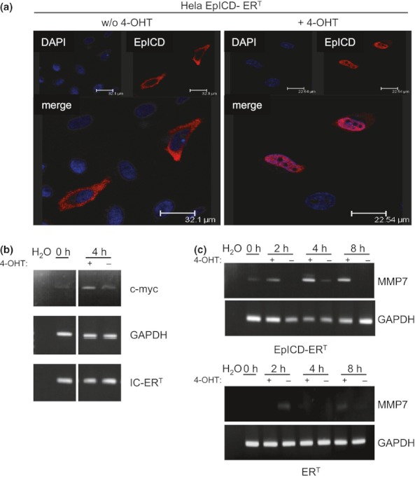
Nuclear translocation of EpICD is essential for transcriptional induction of MMP7. (a) HeLa cells transiently transfected with EpICD-ERT were cultured in the presence or absence of 100 nM 4-hydroxytamoxifen (4-OHT). EpICD-ERT was stained with EpICD-specific antibodies (red) and the localization assessed using confocal laser scanning microscopy. Nuclei were stained with DAPI (blue). Shown are representative images of three independent experiments. (b) At the indicated time points (0 and 4 h post-treatment), mRNAs from non-induced and induced cells were isolated, and the expression of c-myc, GAPDH and IC-ERT was assessed upon RT-PCR with specific primer pairs. Shown are representative results of three independent experiments. H2O served as a negative control. (c) Transient HeLa transfectants expressing ERT or EpICD-ERT were treated with 4-OHT (+) or with solvent only (−), for the indicated time periods (0, 2, 4 and 8 h). mRNA levels of MMP7 and GAPDH were assessed upon RT-PCR with specific primer pairs. Shown are representative results of three independent experiments. H2O served as a negative control.
