Figure 7.

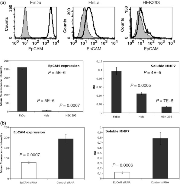
Levels of soluble MMP7 positively correlate with EpCAM expression. (a) Representative histograms of EpCAM FACS stainings in FaDu, HeLa and HEK293 cells are shown. Left panel: Expression of EpCAM was assessed upon flow cytometry with specific antibodies on FaDu, HeLa and HEK293 cells. Shown are mean fluorescence intensity ratios of EpCAM staining vs. control staining with standard deviations. Right panel: Levels of MMP7 in supernatants of FaDu, HeLa and HEK293 cells were measured with an ELISA kit. Soluble MMP7 levels are given as mean relative units (RU) with standard deviations of three independent experiments performed in duplicates. (b) Left panel: FaDu cells were transiently transfected with control siRNA or EpCAM siRNA. After 2 days, EpCAM expression at the cell surface was assessed upon flow cytometry with specific antibodies. Shown are mean fluorescence intensity ratios of EpCAMvs. control stainings with standard deviations from three independent experiments. Right panel: Supernatants from siRNA-treated FaDu cells were collected after 2 days, and levels of soluble MMP7 were measured with an ELISA kit. Soluble MMP7 levels are given as mean relative units (RU) with standard deviations of three independent experiments performed in duplicates. P-values are indicated for each subfigure.
