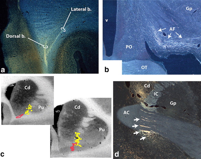
Figure 2.

Photomicrographs and schematics of vmPFC and mOFC pathways. a, vmPFC axons leave the injection site traveling dorsally and divide into the dorsal and lateral bundles. Some dorsal fibers enter the emerging corpus callosum. The medial bundle remains ventral (not illustrated). b, The ventral amygdalofugal pathway carries fibers from the vmPFC to the amygdala. c, Fibers from the vmPFC (red) and mOFC (yellow) enter the IC ventrally and form fascicules within the ventral striatum. d, Photomicrograph illustrating mOFC IC axons embedded within the AC or ventral to it (arrows). Those within the IC terminate in the thalamus; the ventral groups continue to the brainstem. AC, Anterior commissure; AF, ventral amygdalofugal pathway; b., bundle; Cd, caudate nucleus; Gp, globus pallidus; IC, internal capsule; OT, optic tract; PO, preoptic area; Pu, putamen; v, ventricle.
