
Figure 7.
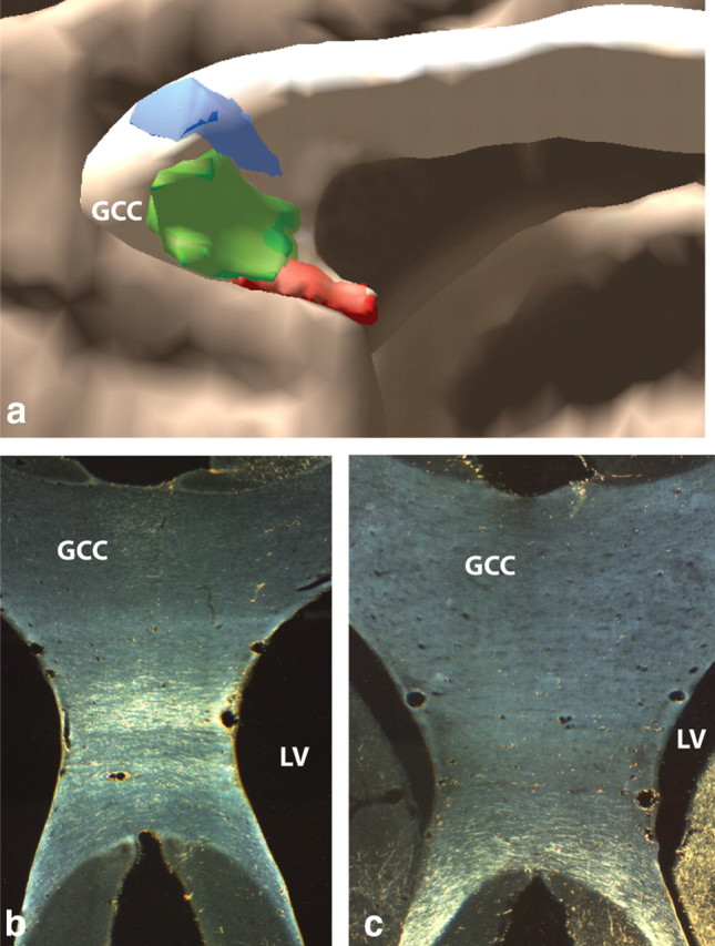
Organization of vPFC pathways in the corpus callosum. a, Sagittal view of 3D model of vPFC pathways in corpus callosum. Fibers originating from more lateral vPFC regions (blue) cross dorsally to fibers from more medial vPFC areas (red). b, Micrograph of cOFC fibers crossing mid-level genu of corpus callosum. c, Micrograph of vmPFC crossing most ventral portion of corpus callosum. GCC, Genu of corpus callosum; LV, lateral ventricle; blue, lOFC; green, cOFC; red, vmPFC.