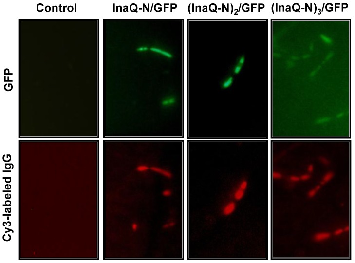
Fig 7.
Micrographs of E. coli JM109/pMB102, JM109/pMB109, and JM109/pMB110 expressing InaQ-N/GFP, (InaQ-N)2/GFP and (InaQ-N)3/GFP, respectively. The panels show the phase-contrast microscopy and fluorescence microscopy images using green and red emission filters. For immunofluorescence microscopy, the cells were treated with anti-GFP monoclonal antibodies, followed with Cy3-conjugated secondary antibodies. E. coli JM109 cells were used as the negative control.

