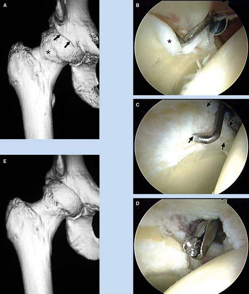
Figure 2.

A 38-year-old woman with progressive pain and loss of motion of the right hip. A, 3-dimensional computed tomography scan illustrates pincer impingement (arrows) and a kissing lesion characterized by osteophyte formation on the femoral head (asterisk). B, as viewed from the anterolateral portal, there is maceration of the anterior labrum (white asterisk) and associated articular delamination (black asterisk). C, debridement of the degenerate labrum exposes the pincer lesion (arrows). D, the pincer lesion is recontoured with a burr. E, a postoperative 3-dimensional computed tomography scan demonstrates the extent of bony recontouring of the acetabulum and the femoral head.
