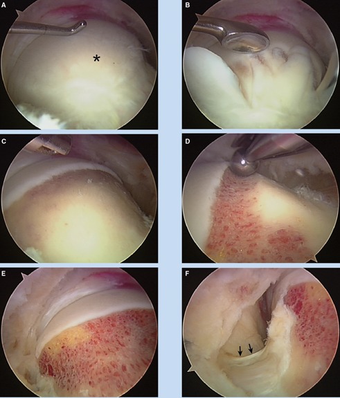
Figure 6.

View from the periphery. A, a cam lesion covered with fibrocartilage (asterisk). B, an arthroscopic curette used to denude the abnormal bone. C, excision area is fully exposed. D, bony resection at the articular margin. E, the completed recontouring. F, lateral view on the base of the neck; the lateral retinacular vessels identified (arrows) and preserved.
