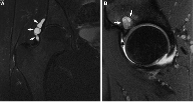
Figure 19.

A, a coronal T2 MRI of a right hip illustrates a paralabral cyst (arrows) pathognomonic of associated labral pathology; B, a sagittal T2-weighted MRI of a right hip demonstrates a subchondral cyst (arrows) indicative of associated articular damage.
