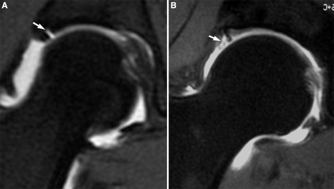
Figure 20.

A, coronal MRA image of a right hip demonstrates contrast separating the lateral acetabulum from the labrum (arrow). Although a labral detachment cannot be ruled out, the smooth margins suggest a normal labral cleft. B, a coronal MRA image of a right hip demonstrates contrast interdigitating within the substance of the lateral labrum (arrow) indicative of true labral pathology.
