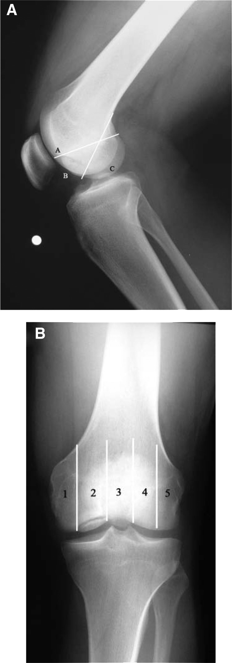
Figure 2.

Anatomic locations of osteochondritis dissecans in the knee. A, lateral radiograph of a 21-year-old man with a BC lesion in the medial femoral condyle; B, anteroposterior radiograph of the same patient shows a grade 2 lesion occupying the weightbearing area of the femoral condyle. Numbering of the 5 anatomic areas begins in the middle side. The condyles are bisected, and area 3 is bounded by the walls of the intercondylar notch.
