
Figure 9.
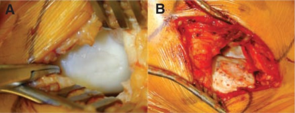
A, intraoperative photo demonstrating a large osteochondritis dissecans lesion located in the medial femoral condyle; exposure was obtained through a medial parapatellar arthrotomy, and the extent of the lesion is determined by direct visualization. B, osteochondritis dissecans lesion following debridement, injection of cultured chondrocytes, and suturing of periosteal graft in place.