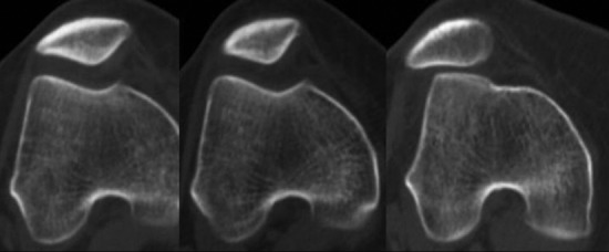
Figure 15.

Computed tomography scan of the patellofemoral joint: from left to right, during 30° of knee flexion with quadriceps contraction, 30° of knee flexion without contraction, and full knee extension without contraction. The image demonstrates lateral subluxation of the patella during only full extension. (Image courtesy of Theodore Miller, MD)
