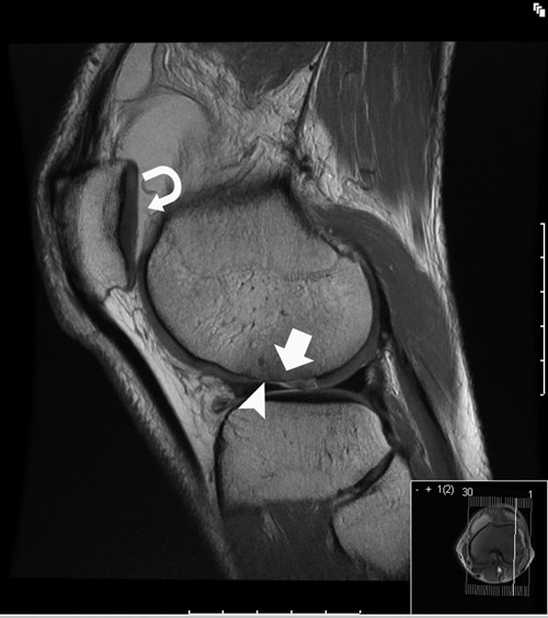
Figure 20.

Sagittal proton density sequence (TR = 4400 milliseconds, TE = 24 milliseconds) demonstrating chondral shear injury of the lateral femoral condyle (arrow) related to patellar dislocation. Note the low signal intensity tidemark (arrowhead) indicative of intact subchondral bone. Proton density–weighted fast spin echo allows differential contrast between articular cartilage and fluid, as seen around the patellofemoral compartment (curved arrow).
