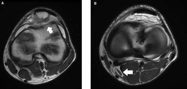
Figure 22.

Axial proton density images (TR = 5858 milliseconds, TE = 25 milliseconds) of the knee in a 13-year-old boy demonstrate (A) traumatic chondral shear of the lateral aspect of the trochlea (arrow) in the setting of a hypoplastic trochlear sulcus, with (B) the loose chondral fragment (arrow) in a popliteal cyst.
