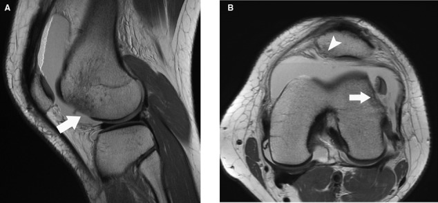
Figure 23.

(A) sagittal proton density sequence (TR = 3750 milliseconds, TE = 25 milliseconds) of the lateral aspect of the knee of a 16-year-old girl demonstrates an osteochondral fracture of the anterior lateral femoral condyle (arrow). (B) axial proton density sequence (TR = 6167 milliseconds, TE = 24 milliseconds) shows the same fracture (arrow), with the osteochondral fragment adjacent to it in the lateral recess of the joint, as well as another osteochondral fracture involving the medial patellar facet (arrowhead). Note the lack of the low signal intensity tidemark, indicative of osteochondral injury.
