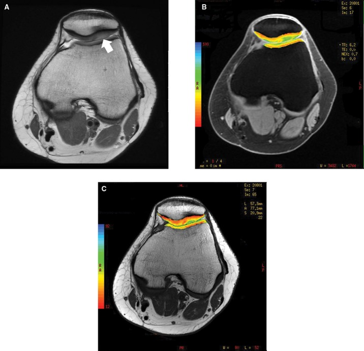
Figure 30.

(A) Axial proton density sequence (TR = 4850 milliseconds, TE = 26 milliseconds) of the knee in a 21-year-old woman who has undergone distal realignment 3 years prior demonstrates loss of the normal gray scale stratification of cartilage along the lateral patellar facet (arrow) with subchondral sclerosis, suggestive of lateral facet overload. T1 rho imaging (B) and T2 mapping (C) demonstrate prolonged relaxation times in the same area, indicating depletion of proteoglycan and abnormal collagen orientation, respectively.
