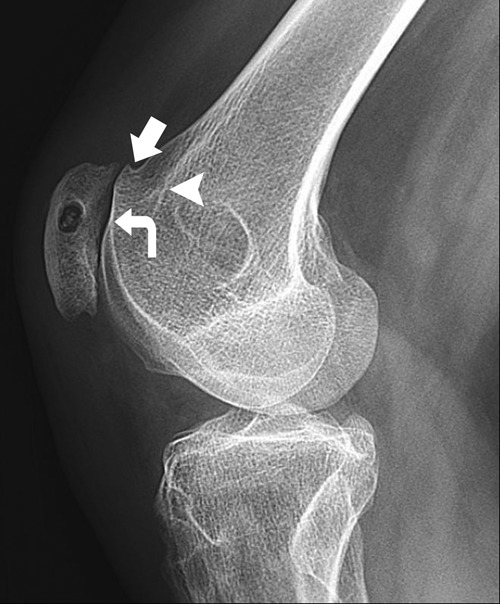
Figure 8.

Lateral knee radiograph of a 32-year-old woman with prior patellar realignment and retinacular reconstruction demonstrating features of trochlear dysplasia, including the “crossing sign” (curved arrow), “supratrochlear spur” (arrow), and “double contour” (arrowhead). Note that this patient had undergone prior medial patellofemoral ligament reconstruction, with the femoral tunnel placed excessively anteriorly.
