
Figure 10.
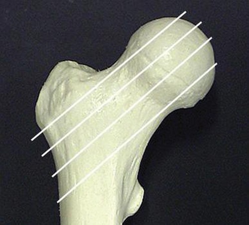
Axial (oblique) images through the femoral neck give the most accurate estimate of femoral anteversion. With the patient in supine position, with symmetric positioning of both lower extremities, and with hips and knees in extension, a coronal-section scout view of pelvis and femur is obtained, parallel to the table. Oblique axial-to-sagittal sections can then be placed parallel to the femoral neck axis, exactly perpendicular to the table. The femoral neck anteversion is determined on the single image, which shows the center of the femoral head and femoral neck.