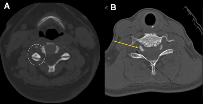
Figure 10.

Axial images of patient in Figure 8 demonstrate the normal configuration (A) and abnormal configuration (B) of the facet joints. A, the so-called hamburger bun configuration. In the circle, note an architecture similar to a hamburger bun. B, there is an uncovering of the facets, or naked facets, with little articulation (black arrow). Note that the top of the bun now points posterior (yellow arrow), instead of the orthotopic anterior.
