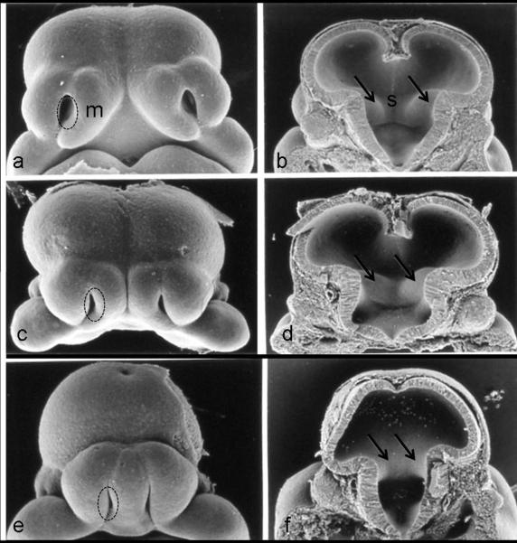
Figure 2.
The face and brain of a normal gestational day (GD) 11 mouse embryo (a, b), as compared to the faces and the brains of two ethanol-exposed embryos (c d; e f ), illustrate the range of defects induced by GD7 ethanol insult. In the ethanol-exposed embryos, loss of midline tissue is notable. Abnormally close apposition of the nostrils (circles) and absence of portions of the medial nasal prominences (m) can be seen in the faces of the ethanol-affected embryos. Similar abnormal proximity of the ganglionic eminences (arrows) can be appreciated (d, f), as can reduction and absence of the septal region (s; d, f). Modified with permission from Sulik KK, Lauder JM, Dehart DB. Brain malformations in prenatal mice following acute maternal ethanol administration. Int J Dev Neurosci 2:203–214, Copyright © 1984 Elsevier and Sulik, Genesis of Alcohol-Induced Craniofacial Dysmorphism Exp Biol Med, 230(6):366-375, Copyright © 2005 The Royal Society of Medicine Press.

