
Fig. 1.
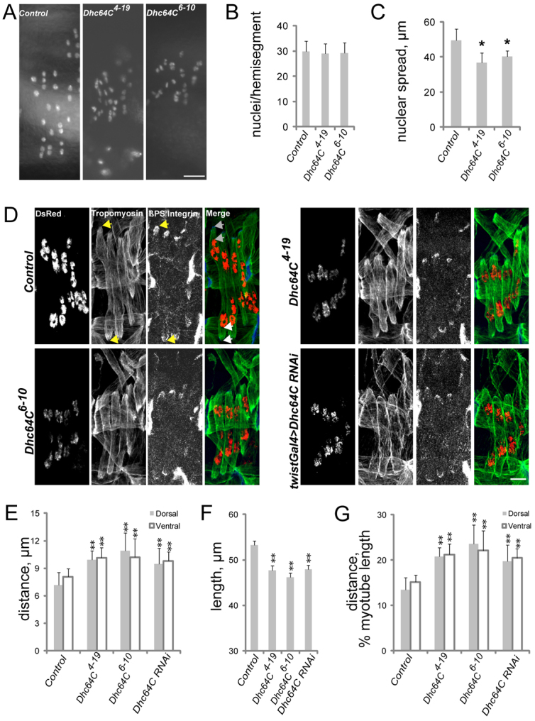
Cytoplasmic Dynein regulates muscle length and myonuclear position. (A) Fluorescence images of apRed nuclei in the lateral transverse (LT) muscles of live stage 17 Drosophila embryos (16.5-20 hours AEL) of the indicated genotypes. (B) The number of nuclei per hemisegment in stage 17 embryos of the indicated genotypes. (C) The distance between the dorsalmost and ventralmost nuclei in stage 17 embryos of the indicated genotypes. (D) Immunofluorescence images of stage 16 (16 hours AEL) embryos of the genotypes noted to the left and antigen listed at the top of the first image. Green, Tropomyosin (muscle); red, DsRed (nuclei); blue, β-PS-Integrin in merge. Arrows in the control panels denote points from which measurements were made for all genotypes. The distance between the points indicated by the yellow arrows in the Tropomyosin and β-PS-Integrin panels was used to determine muscle length. The distance between the pair of gray arrows at the top of the merged image was used to determine the distance between the nuclei and the dorsal pole. The distance between the pair of white arrows at the bottom of the merged image was used to determine the distance between the nuclei and the ventral pole. (E) The shortest distance between the indicated pole of the LT muscles (gray, dorsal; white, ventral) and the nearest cluster of nuclei in stage 16 embryos. (F) LT muscle length in stage 16 embryos. (G) The shortest distance between the indicated pole of the LT muscles (gray, dorsal; white, ventral) and the nearest cluster of nuclei normalized for muscle length in stage 16 embryos of the indicated genotypes. Error bars indicate s.d.; **P<0.01, *P<0.05. Scale bars: 10 μm.