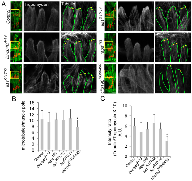
Fig. 7.
Microtubule organization is disrupted in CLIP-190 mutant embryos. (A) Confocal projections of a single hemisegment from stage 16 Drosophila embryos immunostained for Tropomyosin (green) and Tubulin (grayscale). The boxed regions are shown at higher magnification to the right. Yellow arrows indicate individual and/or bundles of microtubules that reach the muscle pole. Scale bars: 10 μm. (B) The number of microtubules within 3 μm of the myofiber pole in the indicated genotypes. (C) The average intensity of Tubulin immunofluorescence in the 3 μm near the myotube pole in the indicated genotypes. Error bars represent s.d.; *P<0.05, compared with control.

