
Fig. 3.
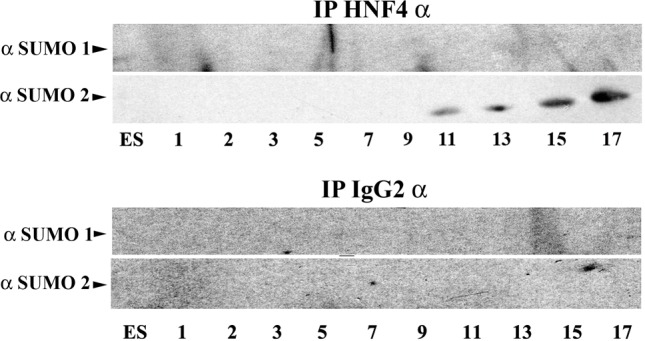
SUMO modification of HNF4α during hepatocellular differentiation. hESCs were differentiated to hepatocytes and samples were harvested at the time points indicated. HNF4α was pulled down using HNF4α antibody covalently crosslinked to Protein-G–Sepharose. An IgG isotype was used as a control throughout. Following incubation and extensive washing, the beads were eluted, separated by SDS-PAGE, western blotted and probed for SUMO1 and SUMO2. We did not detect SUMO1 modification of HNF4α during the differentiation processes. By contrast, we observed an increase in HNF4α SUMO2 modification as differentiation progressed. Our negative control IgG demonstrated that our assay was operating specifically throughout.