
Fig. 6.
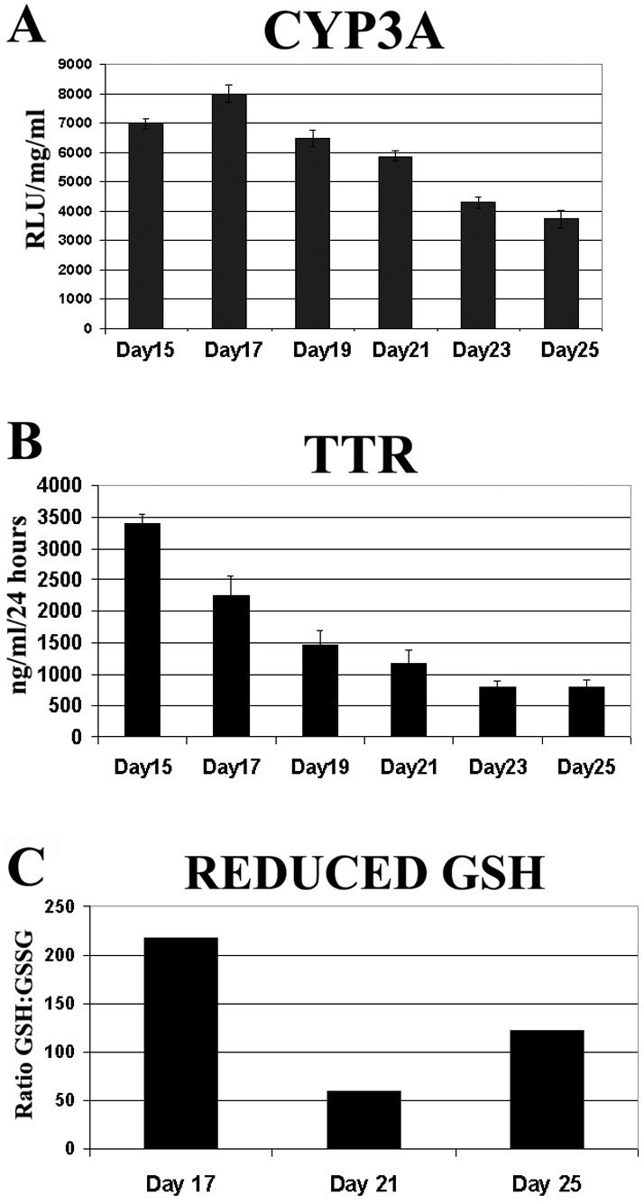
Loss of HNF4α leads to decreased hepatocellular function and oxidative state. (A) Cyp3A metabolic activity of hESC-derived hepatocytes was measured on days 15–25 using CYP3A pGlo substrate. 5 hours post treatment, CYP3A activity was measured on a luminometer. Units of activity are expressed as relative light units (RLU)/mg protein/ml (n = 6). Error bars represent 1 s.d. (B) hESC-derived hepatocytes (days 15–25) were cultured in six-well plates with 1 ml L-15 for 24 hours before culture supernatants were collected and TTR secretion was measured by ELISA. Error bars represent 1 s.d. (C) The redox state of hESC-derived hepatocytes was measured between days 17–25. hESC-derived hepatocytes were incubated with L-15 supplemented with GSH/GSSG pGlo substrate and activity was measured on a luminometer.