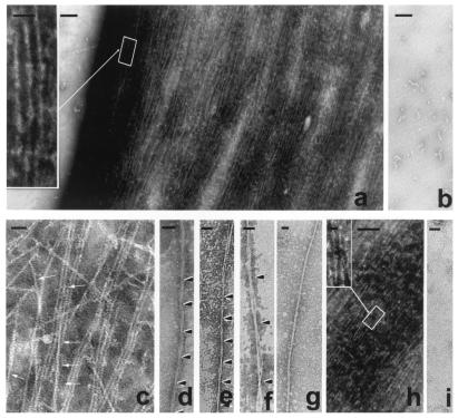
Figure 3.
The formation of tangles of PHF-like and SF from in vitro hyperphosphorylated τ. τ, 0.5 mg/ml, was incubated with rat brain extract in the presence of ATP to induce hyperphosphorylation of τ (a) or incubated with nonhydrolyzable ATP, AMP–PNP, as a control (b). τ hyperphosphorylation (≈12–15 mol of phosphate per mol of protein) induced its self-polymerization into straight and PHF-like (Inset) filaments (a), irrespective of the τ isoform used for the phosphorylation assay. The magnification bars in a and b represent 500 nm and, in the Inset, 50 nm. Intertwining 4-nm filaments generate small PHF-like structures (arrows) as follows: τ4 (c); PHF, τ3L (d); τ protofilaments forming PHF, τ4S (e); τ protofilaments forming bigger PHF, τ4S (f); ≈15-nm straight filament, τ3 (g); mixture of all six τ isoforms hyperphosphorylated (h), and Inset shows a PHF from the tangle formed; and control sample incubated without ATP, τ4S (i). Bars represent 40 nm in c–g and i, 200 nm in h, and 25 nm in Inset.

