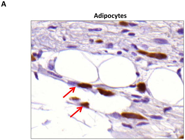
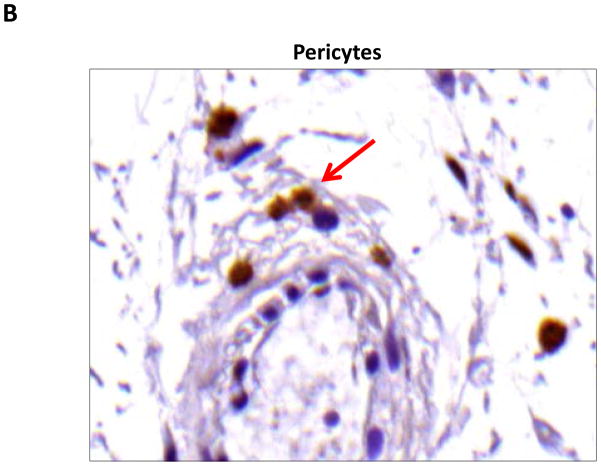
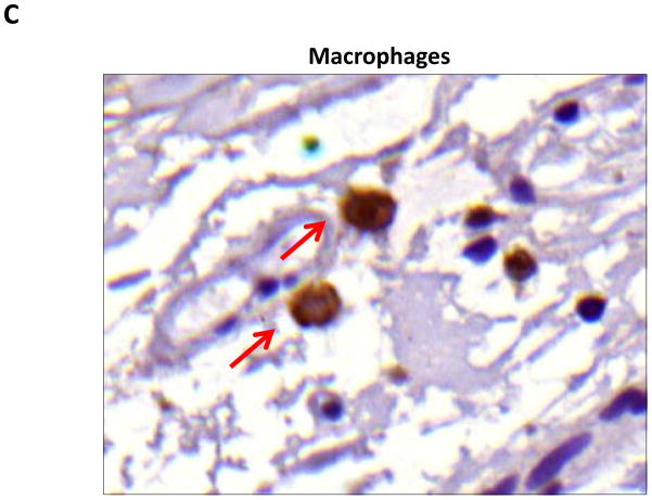
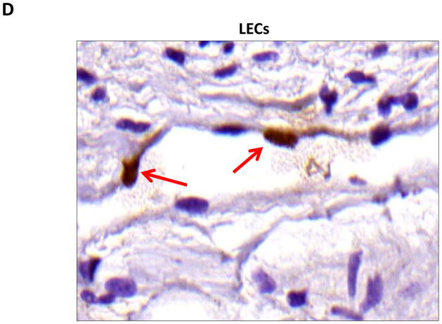
FIGURE 5. PPAR-γ is expressed by a variety of cell types in response to lymphatic fluid stasis.
A–D. PPAR-γ staining (red arrows) of adipocytes (A), pericytes (B), macrophages (C), and lymphatic endothelial cells (LECS; D) shown in representative high power (80X) images of the distal region (D+20) of the tail.


