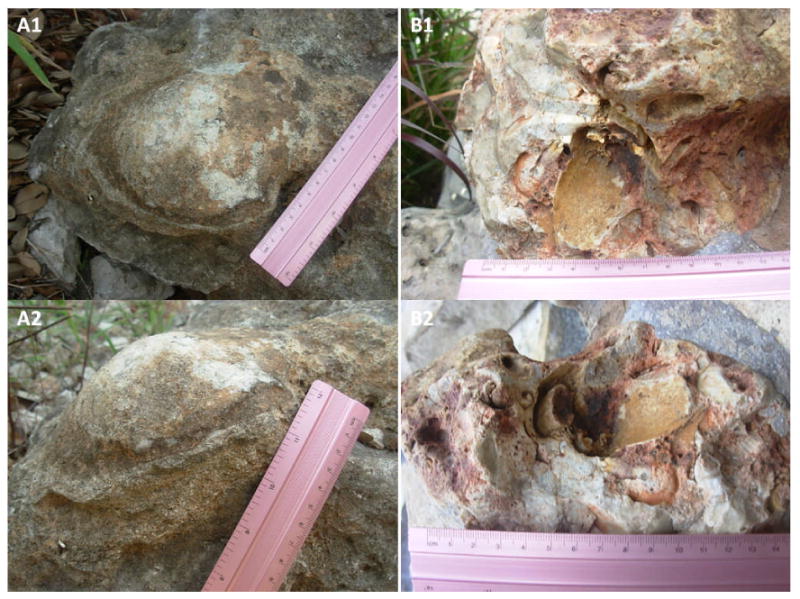
Figure 2.

Images of what appears to be petrified human bones. A1 and A2 show what seems to be a petrified skull covered with a thin and brown peeling layer of what seems to be petrified skin; A2 also shows the detail of a Y-shaped crack in the skull, between the 11 and 11.5 inches mark. B1 and B2 show a petrified tooth and what seems to be bone and medulla remains of a petrified lower leg, the red color within the center of the bigger piece of medulla and of its surroundings indicates a high presence of iron. B1 shows the petrified tooth between the marks of centimeters 2 and 3.5, while B2 shows the bigger petrified medulla between centimeters 4 and 6. These images were taken in Texas.
