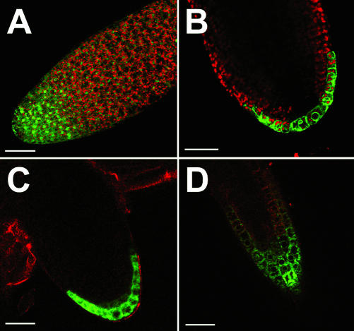
Figure 5.
Comparison of GFP and TM-GFP fluorescence in the tip of an Arabidopsis root (all CLSM). A, Optical section and detection of GFP fluorescence in the root tip an AtSUC3-promoter::GFP embryo. A continuous gradient of GFP fluorescence is seen in the root tip. The soluble, hydrophilic GFP used in this analysis accumulates in the nuclei of all cells and is seen up to 100 μm behind the tip. B, Optical section and detection of TM-GFP fluorescence in the root tip of an AtSUC3-promoter::TM-GFP embryo. The fluorescence is restricted to the outermost cell layer, and TM-GFP labels primarily the cellular membrane systems. C, Optical section and detection of TM-GFP fluorescence in the tip of a very young lateral root of an AtSUC3-promoter::TM-GFP plant. As in the embryo shown in B, TM-GFP fluorescence is restricted to the outermost cell layer. D, Optical section and detection of TM-GFP fluorescence in the tip of a lateral root (slightly older than that shown in C) from an AtSUC3-promoter::TM-GFP plant. At this later stage of lateral root development, TM-GFP fluorescence is seen in the outermost cell layer, but in addition, TM-GFP fluorescence is also detected in the columella cells in the center of the root tip. Scale bars = 40 μm in A, and = 20 μm in B through D.

