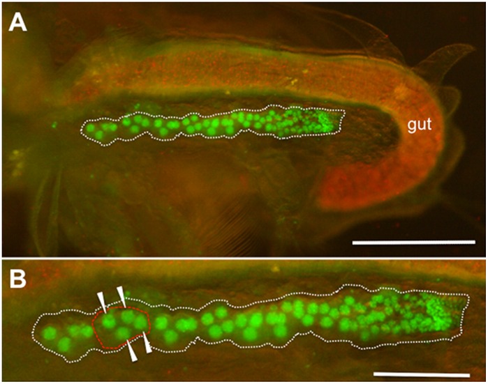
Figure 5. H2B-GFP expression in adult ovaries.
Adult D. magna HG-1 line 2 h after ovulation was incubated in 50% ethanol for 1 min to stop movement and removed the carapace that covered the body. Lateral views with anterior to the left and dorsal upwards. (A) Fluorescence of a thorax and an abdomen in the adult HG-1 line. To show body structure, visible light illumination was also used. Red fluorescence in the gut shows autofluorescence of chlorella. (B) Magnified view of the ovary expressing H2B-GFP. Dotted white and red lines show an ovary and an oocyte cluster comprised of one oocyte and three nurse cells, which are indicated with arrowheads. Smaller nuclei are scattered throughout germarium located at the posterior end of the ovary.

