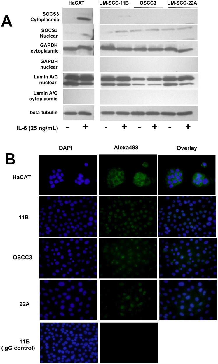
Figure 2. Subcellular distribution of constitutive and IL-6-induced SOCS3 in four different HNSCC cell lines and a non-neoplastic epithelial cell line (HaCAT).
(A) Immunoblot of nuclear and cytoplasmic fractions of protein isolated from three HNSCC cell lines and a non-neoplastic epithelial cell line (HaCAT). HNSCC cell lines that still presented some level of SOCS3 expression and the non-neoplastic HaCAT cells were stimulated with IL-6 (25 ng/mL) for 18 hours. These results support the immunofluorescence findings of a nuclear localization of SOCS3 in HNSCC cells, in contrast to the predominantly cytoplasmic localization in HaCAT cells. Expression of nuclear Lamin A/C and cytoplasmic GAPDH were used as controls for the purity of the different protein fractions, whereas beta-actin was used as a loading control for a mixture of equal concentrations of nuclear and cytoplasmic proteins. (B) The indicated cell lines were plated on chamber slides and treated with IL-6 (25 ng/mL) for 18 h to induce SOCS3 expression. After fixation and permeabilization, cells were stained with a rabbit polyclonal antibody against SOCS3 followed with Alexa488-conjugated anti-rabbit IgG secondary antibody. The nuclei of the cells were counterstained with DAPI. Images from random fields were obtained at 600×magnification and are representative of three independent experiments.

