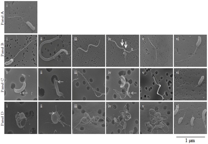
Figure 2. Scanning electron micrographs of V. cholerae strain N16961.
Panel A: Scanning electron microscopy (SEM) image of V. cholerae grown statically overnight at room temperature in L-broth. Panel B: Images obtained with SEM from V. cholerae grown overnight statically at room temperature in FSLW microcosm. Images i to v exhibit diverse V. cholerae morphologies. Image vi obtained after an aliquot of 100 µl of the 24 h old microcosm was transferred to L-broth and incubated statically overnight at room temperature before SEM was performed. Panel C: Images obtained with SEM from V. cholerae persisting statically at room temperature in microcosm for 180 days. Images i through v exhibit different V. cholerae morphologies. Image vi obtained after an aliquot of 100 µl of the 180 days old microcosm was transferred to L-broth and incubated overnight at room temperature before SEM was performed. Panel D: Images obtained with SEM from V. cholerae persisting statically at room temperature in microcosm for 700 days. Images i through v exhibit different V. cholerae morphologies. Image vi obtained after an aliquot of 100 µl of the 700-day old microcosm was transferred to L-broth and incubated overnight at room temperature before SEM was performed. (scale bar, 1 µm; thick arrows indicate evidence of cell division; thin arrows indicate bud and OMVs formation).

