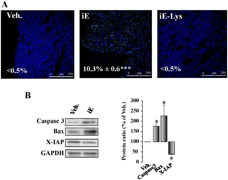
Figure 4. Selective stimulation of NOD1 induces cardiac apoptosis.
Animals received i.p. 150 µg/day of iEDAP (iE), 150 µg/day of iE-Lys or vehicle for 2 weeks. (A) TUNEL staining of cells undergoing apoptosis in cardiac tissue sections from vehicle, iE or iE-Lys-treated mice. Light transmission of the preparations indicated that apoptotic cells were predominantly cardiomyocytes. Representative images of TUNEL positive (green) and DAPI (blue) staining (magnification x 40). The percentages of positive TUNEL cells are indicated in the images. (B) Caspase 3, Bax and X-IAP levels determined by Western blot from hearts of vehicle and iE mice. Left panel shows the histograms representing the mean±SEM vs. vehicle (100%). *p<0.05, ***p<0.001 vs. vehicle; n = 4–6 animals.

