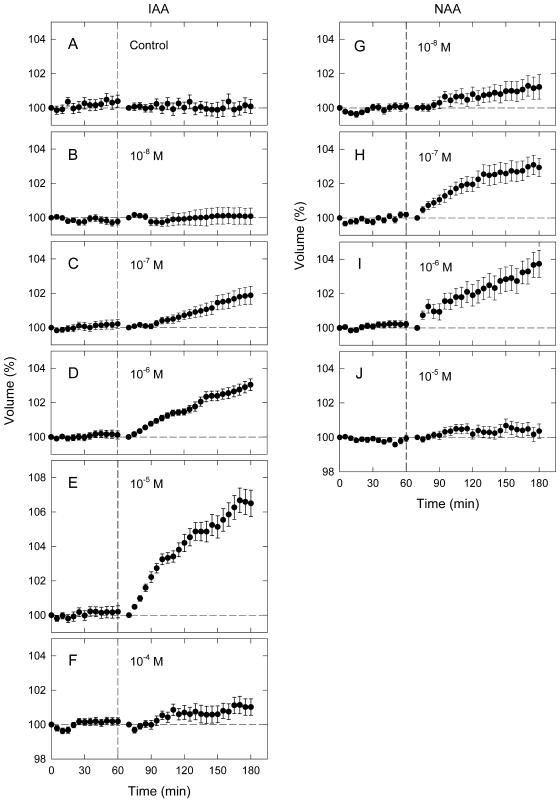
Figure 1.
Auxin-induced swelling of pea epidermal protoplasts. Photographic recording of protoplast images was initiated at time zero, which was 30 min after the onset of incubation on the microscope stage. At the vertical dashed line (immediately after obtaining protoplast images at 60 min) a solution containing indole-3-acetic acid (IAA; B–F) or 1-naphthalene acetic acid (NAA; G–J) in bathing medium was added to the concentrations indicated. The control (A) was obtained by adding bathing medium alone. The relative volume of each protoplast was calculated as the percentage of the initial volume, which was the volume at time zero (the time course before auxin application) or 70 min (the time course after auxin application). The means from 98 to 144 protoplasts (A–F; three experiments) or 65 to 82 protoplasts (G–J; two experiments) are shown. Vertical bar = se.

