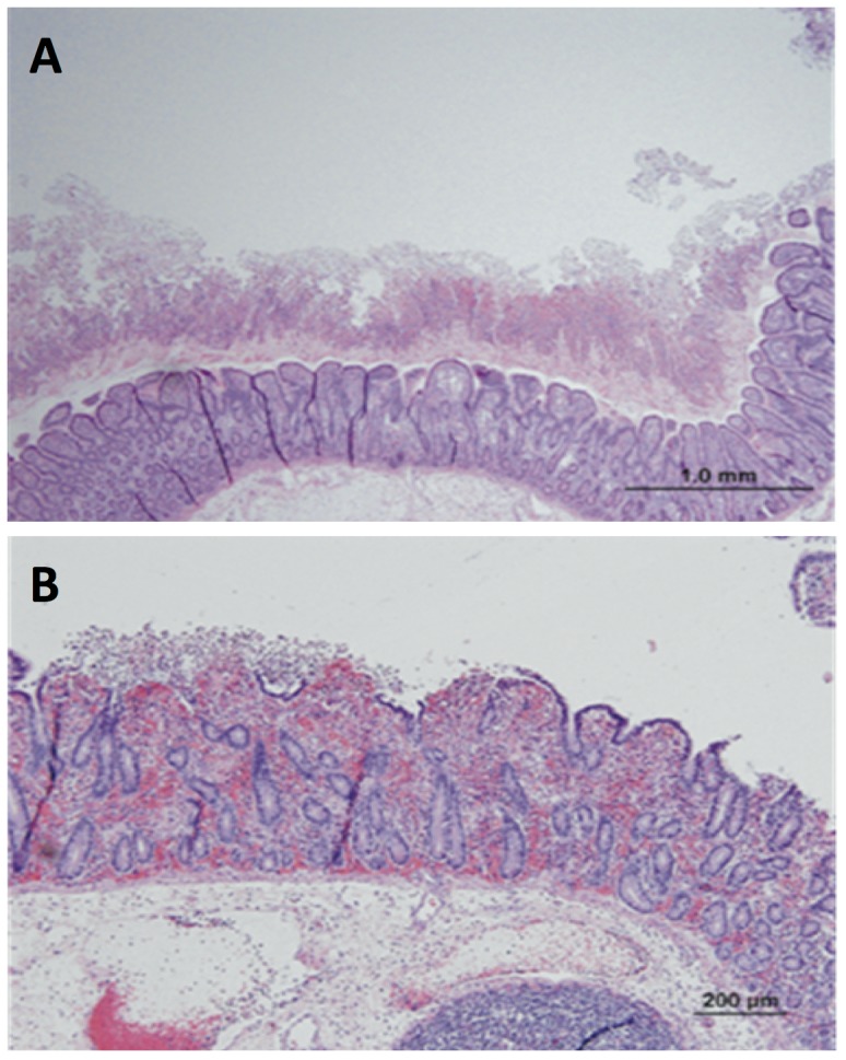
Figure 1. Microscopic sections of the ileal loops representing pseudomembrane formation due to the infection of C. difficile under the experimental conditions.

(A) The mucosal surface is covered by a thick layer of fibrinopurulent exudate (‘pseudomembrane’). Note the extensive submucosal edema. (B) Focal erosion of the surface enterocytes is accompanied by exudation of fibrin and neutrophils into the lumen of the intestine.
