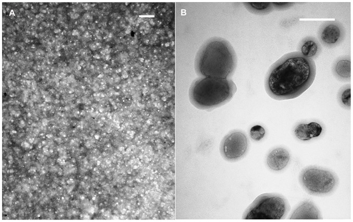
Figure 3. Transmission Electron Micrograph (TEM) of h9h5-vesicles.
These peptides self-assemble into nano-sized vesicles. TEM images of (A) a concentrated and (B) a diluted sample of the peptide mixture, stained with 5% phosphotungstic acid and Osmium tetroxide (OsO4) vapors, respectively (200 nm scale bar).

