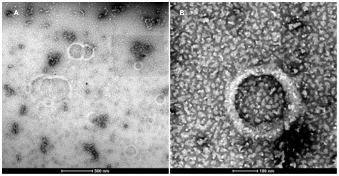
Figure 4. Scanning Transmission Electron Micrograph (STEM).
Vesicles were prepared with 30% CH3-Hg label in both the h5 and h9 peptides at 0.1 mM concentration were negatively stained using a multi isotope 2% Uranyl acetate (Uranium bis(acetato)-O)dioxo-dihydrate) aqueous solution. The images were captured using annular dark field mode was then inverted to produce the final image.

