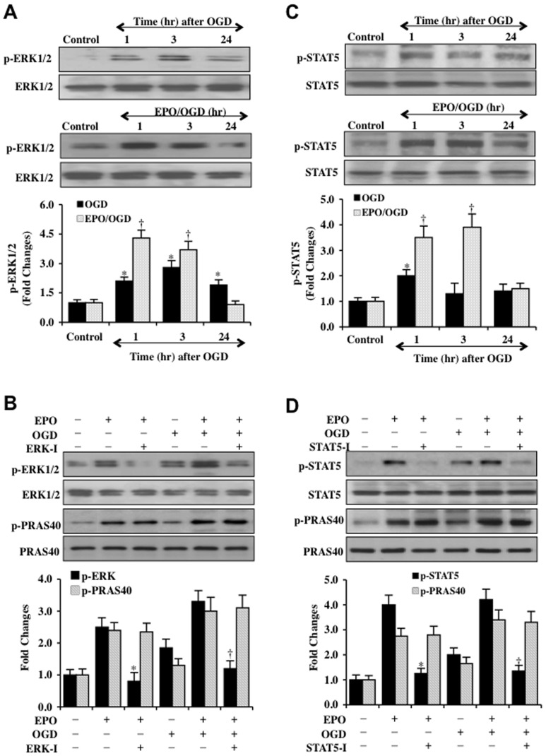
Figure 7. EPO pathways of ERK 1/2 and STAT5 do not alter phosphorylation of PRAS40.
(A) Western blot analysis for phosphorylated (p-) ERK 1/2 (p-ERK 1/2, Thr202/Tyr204) in SH-SY5Y cells was performed at 1, 3, or 24 hours (hr) following OGD. EPO (10 ng/ml) that was applied to cell cultures 1 hour prior to OGD significantly increased p-ERK 1/2 expression at 1 and 3 hours following OGD (*P<0.01 vs. Control; † P<0.01 vs. OGD at corresponding time points). (B) EPO (10 ng/ml) that was applied to SH-SY5Y cells significantly increased the expression of phosphorylated (p-) p-ERK 1/2 either alone or during OGD exposure 3 hours later. Expression of p-ERK 1/2 was significantly limited during the application of the ERK inhibitor (ERK-I, 100 μM) applied to cultures 30 min prior to EPO administration. Inhibition of ERK 1/2 did not alter the ability of EPO to significantly phosphorylate (p-) p-PRAS40 with or without OGD exposure (*P<0.01 vs. EPO; † P<0.01 vs. EPO/OGD). (C) Western blot analysis for phosphorylated (p-) STAT5 (p-STAT5, Tyr694) in SH-SY5Y cells was performed at 1, 3, or 24 hours (hr) following OGD. EPO (10 ng/ml) that was applied to cell cultures 1 hour prior to OGD significantly increased p-STAT5 expression at 1 and 3 hours following OGD (*P<0.01 vs. Control; † P<0.01 vs. OGD at corresponding time points). (D) EPO (10 ng/ml) that was applied to SH-SY5Y cells significantly increased the expression of phosphorylated (p-) p-STAT5 either alone or during OGD exposure 3 hours later. Expression of p-STAT5 was significantly limited during the application of the STAT5 inhibitor (STAT5-I, 100 μM) applied to cultures 30 min prior to EPO administration. Inhibition of STAT5-I, 100 μM did not alter the ability of EPO to significantly phosphorylate (p-) p-PRAS40 with or without OGD exposure (*P<0.01 vs. EPO; † P<0.01 vs. EPO/OGD). In all cases above, each data point represents the mean and SD from 3 experiments.

