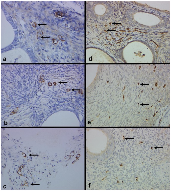
Figure 7. Microvessel density (MVD) of implants stained with α-SMA (a–c) and vWF (d–f) in control, PEDF-5 and PEDF-1 groups (Original magnification ×400).
There is no difference between control (a) and PEDF-treated groups (b, c) in α-SMA labeled MVD, while MVD labeled by vWF in the PEDF-1 group (f) is significantly reduced compared to control group (d).

