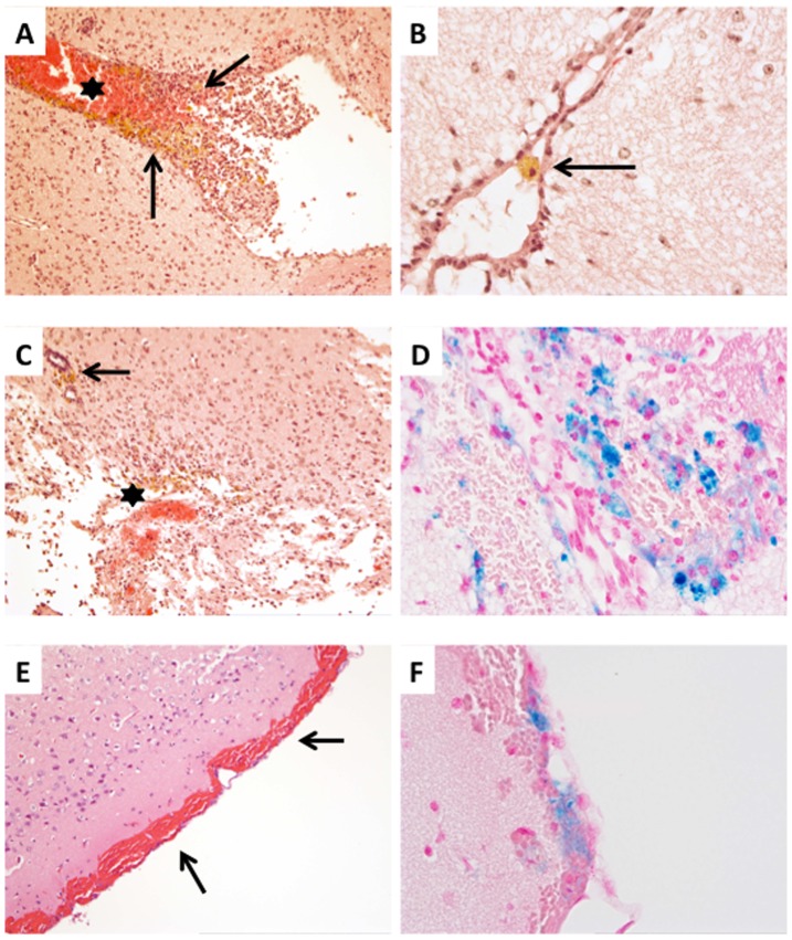
Figure 7. Hydrocephalus in JAM-C−/− C57BL/6 mice is accompanied by hemorrhages.
A: Hematoxylin and eosin staining showing signs of fresh (asterisk) and elder (arrows) bleedings within the III. ventricle. Elder bleedings consisted of erythrophages, hemosiderophages and also haematoidin pigment, the latter indicating bleedings older than 8 days (original magnification: 10x). B: Signs of elder bleedings were also observed within the aqueduct (arrow indicating hemosiderophage; HE stain; original magnification: 40x). C: Fresh and elder hemorrhages were also seen in the perivascular spaces (arrow) and within the CNS tissue (asterisk) (HE stain; original magnification 10x). D: Iron staining indicating the presence of abundant intraparenchymal iron-loaden macrophages (original magnification: 40x). E, F: Subarachnoidal hemorrhages presenting with areas of fresh (E; arrows; HE staining; original magnification: 10x) and elder bleedings (F; iron staining; original magnification: 40x).

