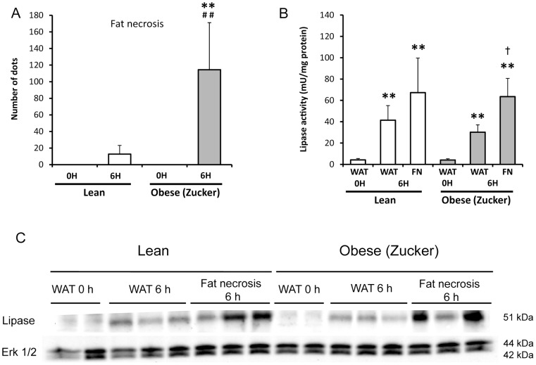
Figure 7. Fat necrosis and pancreatic lipase in abdominal white adipose tissue from lean and obese rats with acute pancreatitis.
Macroscopic quantification of fat necrosis in lean and obese (Zucker) rats at 0 and 6 hours after induction of acute pancreatitis (A). Presence of pancratic lipase in adipose tissue in acute pancreatitis is illustrated by the increase of pancreatic lipase activity in white adipose tissue (WAT) and fat necrosis (FN) (B). A representative image of the presence of pancreatic lipase in WAT and FN is shown (C). Erk 1/2 was used as loading control. The number of rats per group was 8–10 for A and B, and 6–9 for C. The statistical difference is indicated as follows: ** P<0.01 vs. time “0”. ## P<0.01 obese vs. lean in the same conditions. † P<0.05 fat necrosis vs. WAT at 6 hours in obese rats.

