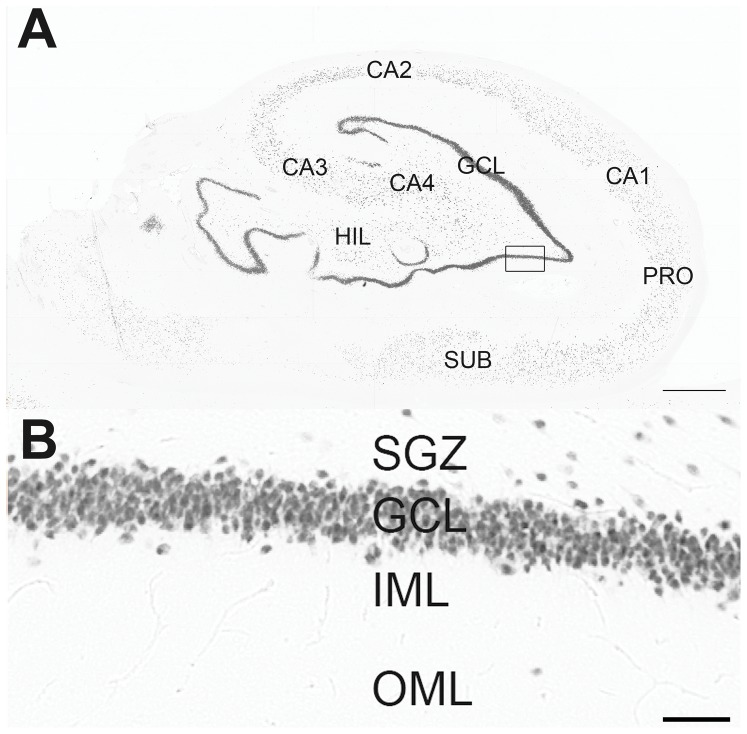
Figure 1. Subfields in the hippocampal formation under NeuN immunohistochemistry.
In A can be seen: the granule cell layer of fascia dentata (GCL, composed by granular neurons) and the hilus (HIL, composed by several types of interneurons); pyramidal neuronal layers of the hippocampus (CA4-CA1); the subicular formation, composed by prosubiculum (PRO) and subiculum (SUB). In B, a higher magnification of the fascia dentate (marked as a black square in A), composed by subgranule zone (SGZ), granule cell layer (GCL), inner molecular layer (IML) and outer molecular layer (OML). Bar in A indicates 1 millimeter and in B indicates 50 micrometers.

