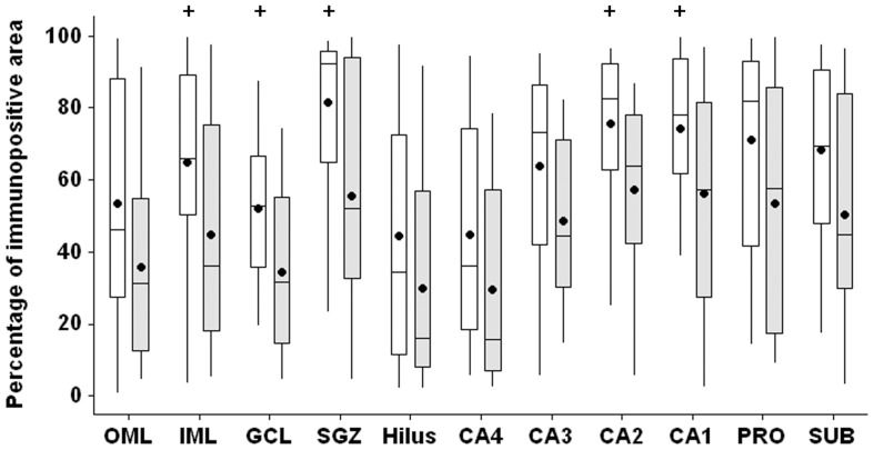
Figure 11. MT-I/II immunopositive area in MTLE patients without and with secondary generalized seizures.
Patients without secondary generalization (white boxplots) present increased MT-I/II immunopositivity (p<0.05) in the inner molecular layer (IML), granule cell layer (GCL), subgranule zone (SGZ), CA2 and CA1, when compared with patients that present secondary generalization (light gray boxplots). The + indicates difference between the groups.

