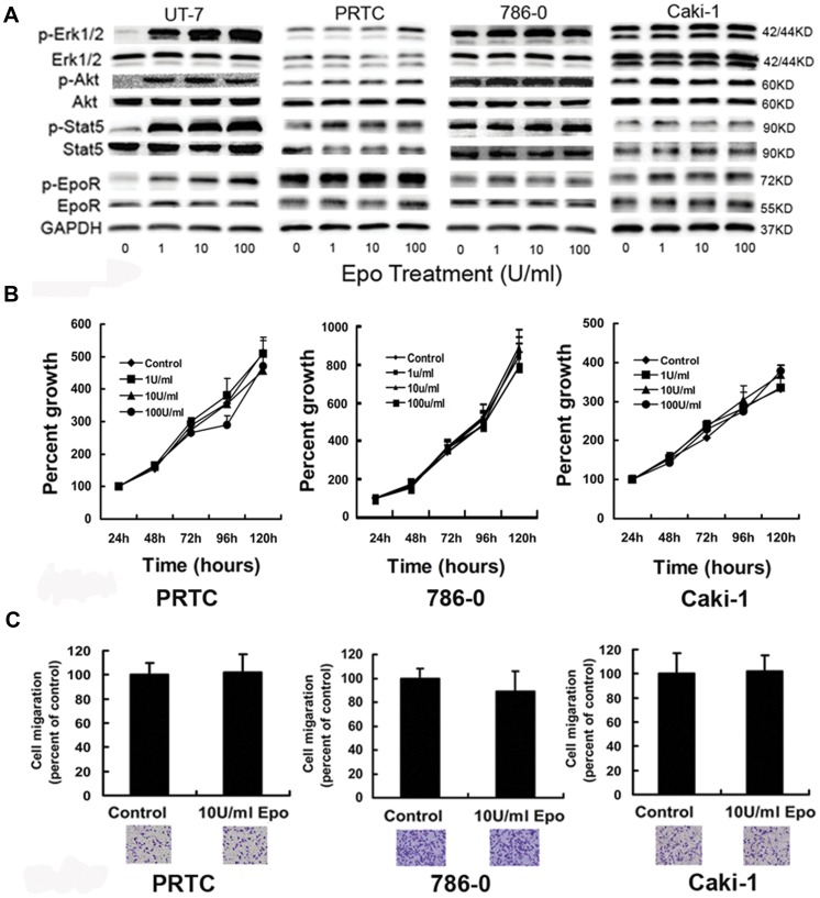
Figure 2. Exogenous Epo shows no biological effect on signaling molecules, proliferation, and invasion ability in RCC cells.
(A) RCC cells were treated with 0–100 U/ml Epo for 5 min after serum starvation for 24 hours. Compared with Epo-dependent UT-7 cells, the phosphorylation of EpoR, STAT5, Akt and Erk1/2 in PRTC, 786-0 and Caki-1 cells increased insignificantly, if any, after rhEpo stimulations. Moreover, RCC cells expressed higher levels of baseline phosphorylation of all examined EpoR signaling proteins than UT-7 cells in the absence of exogenous Epo. In UT-7 cells, p-EpoR, p-STAT5, p-Erk1/2 and p-Akt were at lower levels before Epo stimulation, and increased significantly at 1 U/ml Epo. Experiments were done in triplicate with similar results. (B) PRTC, 786-0 cells and Caki-1 cells were cultured in media containing 0, 1, 10 and 100 U/ml Epo after serum starvation for 24 hours. Viable cells were evaluated after various incubation periods by modified MTT assay. Exogenous Epo had little effect on proliferation of RCC cells (6 wells for one sample, and experiments in triplicate; P>0.05 by one-way ANOVA). (C) Transwell tests for invasion ability of PRTC, 786-0 cells and Caki-1 cells. Upper chamber: 2×10⁴ RCC cells in serum-free medium with 0-100 U/ml concentrations of Epo; lower chamber: medium containing 10% FBS. Cells were stained and counted after incubation for 36 hours. Error bar represents mean ± SD (n = 3). There is no difference between RCC cells with Epo and those without Epo in medium (only showing 10 U/ml groups).

