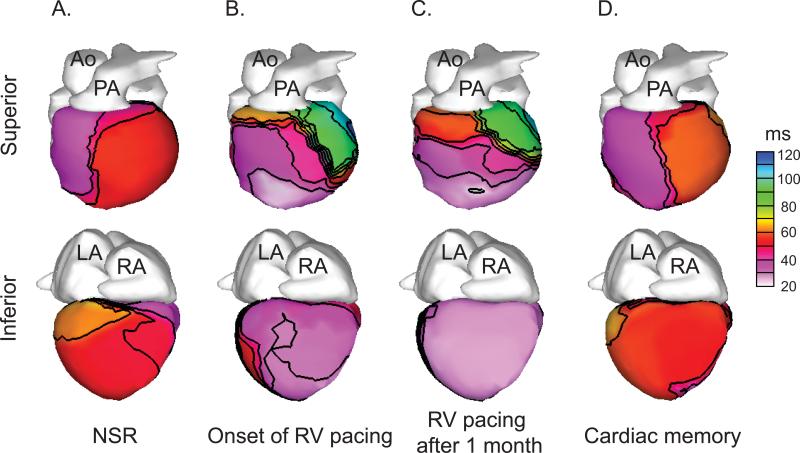
Figure 2.
Epicardial activation times do not exhibit persistent changes after a period of RV pacing. Representative epicardial activation time isochrones are shown. A. Epicardial activation during normal sinus rhythm exhibits a broad area of early activation in the RV. B-C. Epicardial activation during RV pacing exhibits a site of early activation over the apical RV followed by delayed activation of the basal LV. D. Epicardial activation after restoration of NSR is similar to the baseline activation pattern (in A). Ao = aorta; PA = pulmonary artery; LA = left atrium; RA = right atrium; NSR = normal sinus rhythm. Time values (in ms) are in reference to the onset of the body-surface QRS.

