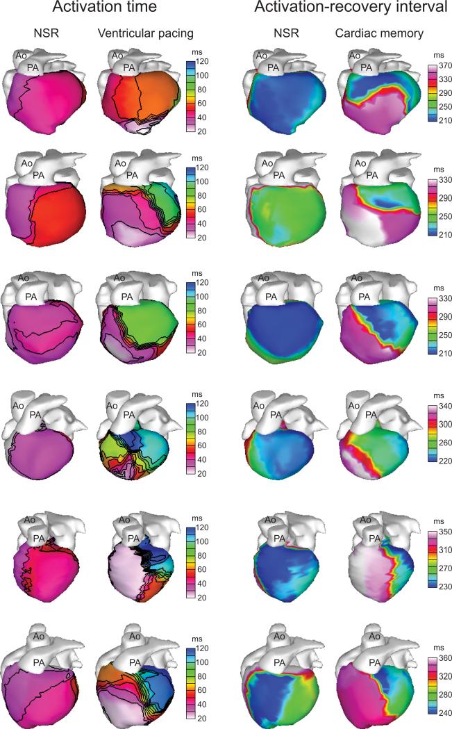
Figure 4.
The region of ARI prolongation in all subjects corresponds to the region of early pacemaker-induced activation. Each row represents an individual subject in the study. For each subject, the left two columns show isochrones of epicardial activation during NSR and RV pacing to demonstrate the location of early pacemaker-induced activation. The right two columns show epicardial ARI maps during NSR and cardiac memory, demonstrating a similar pattern of ARI prolongation during cardiac memory to the early activation during RV pacing.

